LSU, Tulane Researchers Synthesize Ultrasmall Nanoshells for Improved Biomedical Imaging
 September 21, 2023
BATON ROUGE, LA – Researchers from LSU’s Cain Department of Chemical Engineering and Tulane University’s Biomedical Engineering Department synthesized sub-100 nm gold nanoshells, i.e., particles with silica cores covered by a thin gold shell, and used them to improve the penetration depth of photoacoustic imaging. Gold nanoshells are promising candidates for biomedical imaging and cancer therapy, but synthetic challenges have limited their diameters to greater than 100 nm for more than 20 years. LSU chemical engineers synthesized 62 nm nanoshells and collaborated with Tulane biomedical engineers to demonstrate a 50% improvement in the photoacoustic imaging depth compared to current state-of-the-art nanoshells.
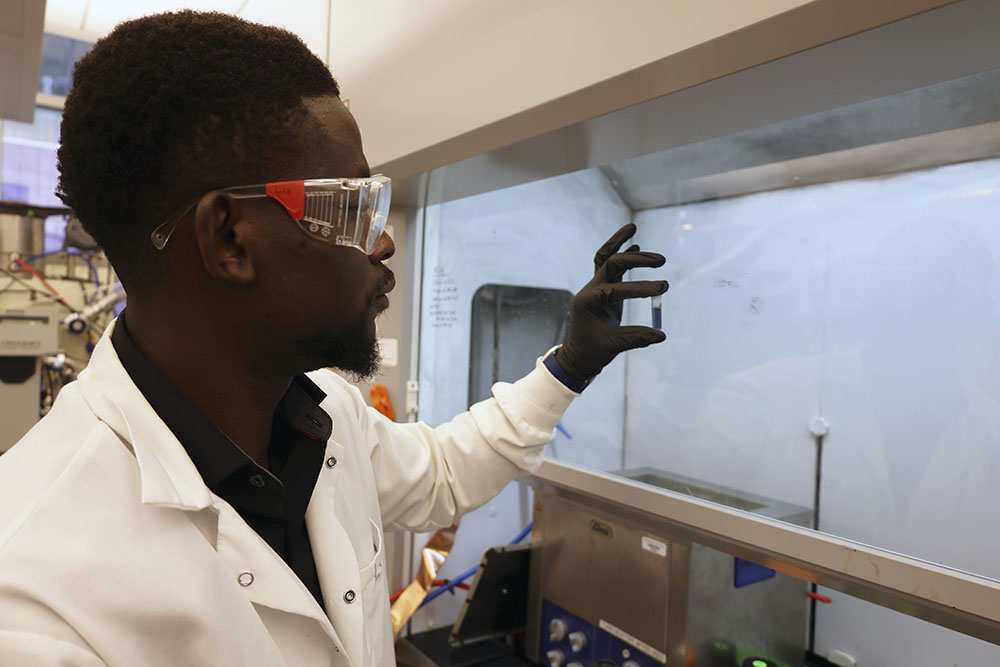
The research team working on the project consists of LSU Chemical Engineering (ChE) Ph.D. student Luis Manuel, LSU ChE Associate Professor Kevin McPeak, Tulane University Biomedical Engineering Ph.D. student Vinoin Devpaul Vincely, and Tulane University Biomedical Engineering Associate Professor Carolyn Bayer. The work was recently published in the journal Nano Letters, titled, “Monodisperse Sub-100 nm Au Nanoshells for Low-Fluence Deep-Tissue Photoacoustic Imaging”.
Photoacoustic imaging is a relatively new, non-invasive biomedical imaging technique that uses light to generate sound waves in human tissue and ultimately obtain internal structure and chemical information about the human body. During photoacoustic imaging, sound waves are detected by a transducer, just like ultrasound imaging, a common method used to image pregnancies. Unlike ultrasound imaging, photoacoustic imaging has micron-scale spatial resolution, which allows it to generate higher-quality images of tissue and organs. The absorption of light by the body and subsequent generation of heat and, ultimately, sound waves is sensitive to the chemical environment in the body. This chemical information can simultaneously be acquired along with structural information. Still, not all biomarkers in the body absorb light, and it’s often challenging to separate the background signal from the area of interest.
“Introducing nanoparticles into the body can extend the functionality of photoacoustic imaging by allowing us to target certain biomarkers that do not absorb light,” McPeak said. “We show that ultra-small gold nanoshells can help overcome these challenges by minimizing scattering losses and efficiently absorbing near-infrared light and generating heat. Large nanoshells (greater than 150 nm in diameter) have been employed since the 1990s, and clinical trials have been undertaken. However, these larger nanoshells scatter light stronger than they absorb, resulting in a weak photoacoustic imaging response. Synthesis methods over the past 20 years for reducing the size of nanoshells have failed. We overcome these synthetic challenges to make nanoshells down to 60 nm in diameter that strongly absorb light and provide excellent photoacoustic imaging contrast agents.”
You can read the research team’s full paper by clicking here.
Like us on Facebook (@lsuengineering) or follow us on Twitter and Instagram (@lsuengineering).
###
Contact: Libby Haydel
Communications Manager
225-578-4840
ehaydel1@lsu.edu