How Having Our Own High-Resolution Avatar Could Transform Healthcare—in Cancer Clinics and in Space
January 18, 2022
Students in the LSU SpaRTAN medical physics lab are using technology to simulate biology and biological processes, such as a person’s whole-body response to radiation based on the break-down of individual cells. And the result is beautiful.
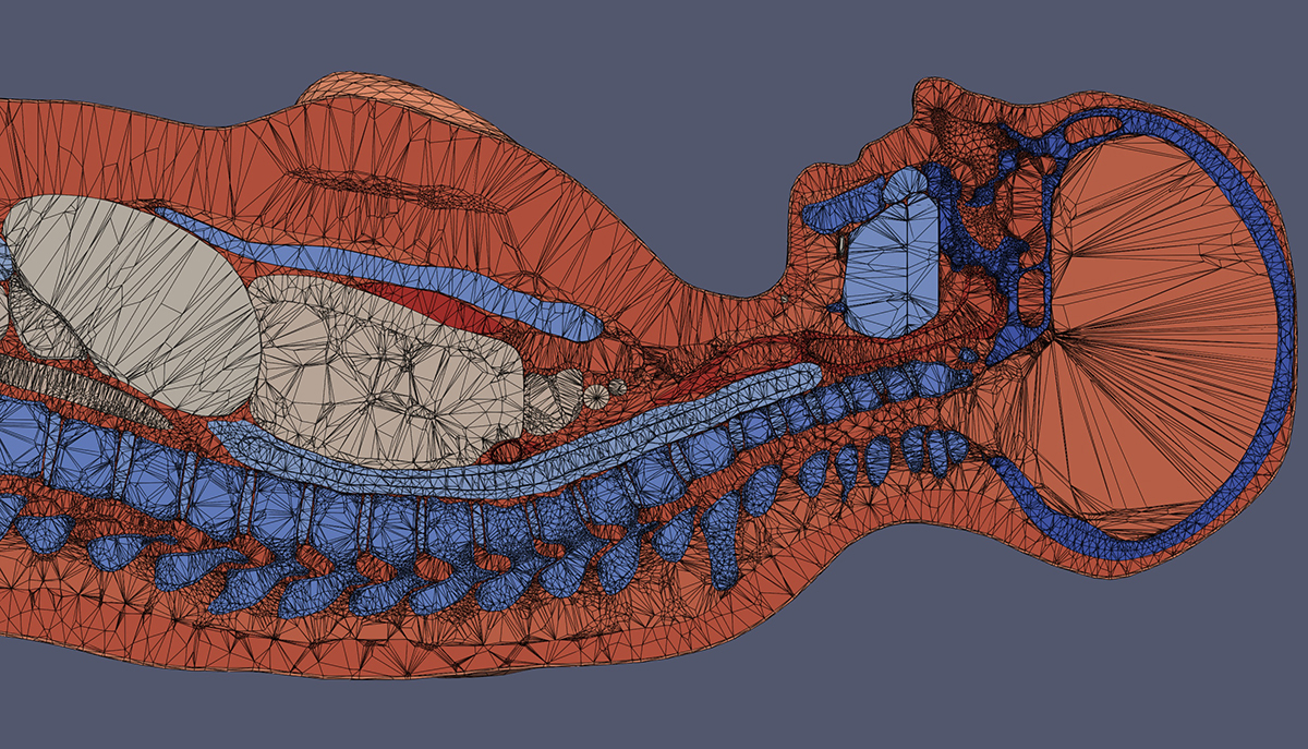
The computational human phantoms developed in the LSU SpaRTAN lab are high-resolution renderings of human bodies, including blood, bone, organs, and tissues. One of the advantages of visualizing the whole body in 3D is to be able to safely study its dynamic response, interactions between tissues and organs, and slight differences between them.
– Megan Chesal / LSU
Baton Rouge—On a day-to-day basis, LSU medical physics graduate student Megan Chesal might not feel like Leonardo da Vinci as she sits at her computer in her two-bedroom apartment in Baton Rouge, trying to understand the complex workings of the human body by creating highly intricate drawings. But she’s nevertheless following in his footsteps by using technology to describe biology.
Chesal develops human phantoms, which are computational 3D replicas of entire bodies. These virtual alter egos or avatars can be used for medical research to help predict outcomes without having to experiment on living beings. Chesal’s longterm goal is for her research to help in the fight against cancer, which can be treated with radiation. More immediately, however, her focus is on space radiation and its effects on the human cardiovascular system. Space radiation effects, to both soft and condensed matters, is the primary focus of the LSU SpaRTAN lab, which she helps manage.
“Radiation has the power to disrupt or kill cells—whether those are healthy cells, or cancer cells.”
Chesal and her fellow students are all studying radiation effects under the supervision of Jeffery Chancellor, assistant professor in the LSU Department of Physics & Astronomy. The students research radiation in space, working on ways to protect astronauts and equipment from harmful background radiation, while some also look at it as a positive, in the context of medicine. In either scenario, radiation has the power to disrupt or kill cells—whether those are healthy cells, or cancer cells. The phantoms Chesal is developing can be used to study space radiation deposition and topology throughout the body—she’s a current Louisiana Space Grant graduate research fellow—as well as for other purposes, such as predicting and visualizing the distribution and accumulation of pretty much any matter or medication that enters the body.
“Our lab is unique in that we conduct research that can impact both space travel and cancer care, but helping to treat cancer was the reason I went into medical physics and it’s still something I want to do and feel passionate about,” Chesal said.
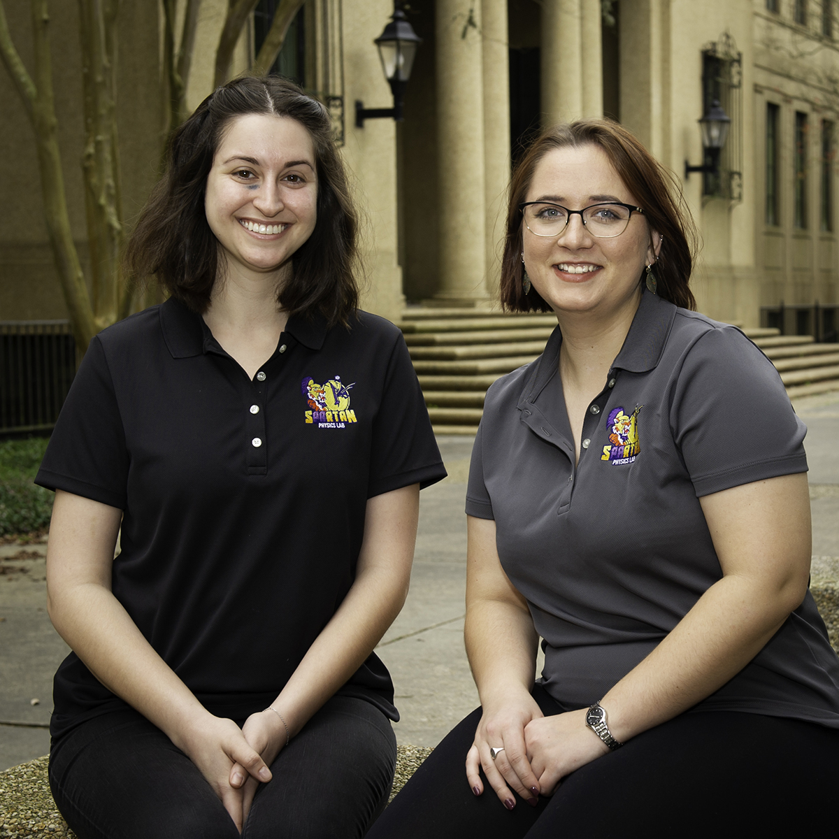
By combining physics, biology, medicine, high-performance computing, and art, Megan Chesal (right) and Nousha Afshari (left) are working to create human phantoms—virtual avatars—that are both anatomically and functionally accurate to help predict and visualize individual outcomes when astronauts and cancer patients are exposed to radiation. (Radiation and chemotherapy are the most common treatments for malign tumors.)
– Elsa Hahne / LSU
The process of building a human phantom starts with a scan, such as an X-ray or magnetic resonance imaging (MRI). Through advanced computational techniques, that data is then translated into virtual 3D. Chesal’s daily challenge is to make these renderings increasingly accurate and more detailed. She spends a lot of time thinking about how to computationally describe and delineate thin and particularly radiosensitive tissues, like oral mucosa and intestinal linings. When she first started building human phantoms, she was working with voxels, or volume pixels, and has since moved on to tetrahedral models. From a computational as well as anatomical standpoint, it makes a big difference whether you’re working with squares/cubes or triangles/pyramids. The latter is much easier to scale and better at describing smooth surfaces. If you were creating a digital drawing of a heart, for example, and then had to draw a much bigger heart, you would need to add a lot more cubes. You couldn’t just make the cubes bigger—it would be like building with oversized Legos, and jagged. Pyramids, on the other hand, can stretch, without as much distortion.
“Imagine, a billion cells responding. You can’t run a simulation like that on a laptop.”
Megan Chesal
“Working with voxels, which are like little boxes, there were regions of the body I couldn’t even describe,” Chesal said. “You get these weird, stairstep surfaces instead of thin, smooth layers, and that can cause problems in calculating something like radiation dose, because no matter how you try, you’ve either included some part of the air, or accidentally taken out parts of the body. Resolution becomes extremely important, but then the computation also becomes massive. Imagine, a billion cells responding. You can’t run a simulation like that on a laptop.”
While Chesal works with tetrahedrons to add information about smaller and smaller details, making her phantoms more anatomically correct, she also collaborates closely with another graduate student in the SpaRTAN lab, Nousha Afshari, who is studying radiation effects on microscopic cells all the way up to macroscopic tissues and organs, sort of in the opposite direction—from the super-small to the larger. (Afshari recently visited SpaceX in California along with another student in their lab, Jared Taylor, to present SpaRTAN research.) Their mutual goal is to have their research meet and then overlap, so the anatomically correct phantoms become functionally correct.
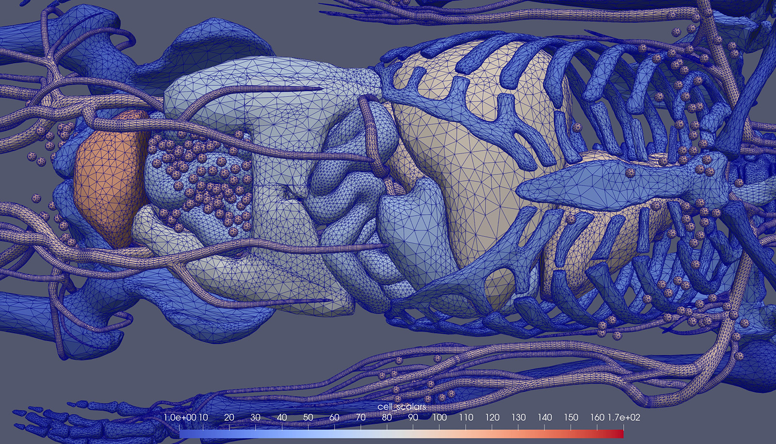
Computational human phantoms for medical research share a common ancestor with the type of models used for video games and virtual production to make fictional characters move in accordance with how their joints (should) work, muscle mass, and weight.
– Megan Chesal / LSU
Before joining LSU, Afshari worked as a medical scribe at Our Lady of the Lake Voice Center in the same building as Mary Bird Perkins Cancer Center, in Baton Rouge. There, she became aware of how difficult it can be to predict outcomes for individual cancer patients—even seemingly similar patients with the same kind of tumor.
“There are a ton of factors that influence how somebody responds to radiation as well as cancer,” Afshari said. “Damage is happening at a microscopic level, and yet most of our tools make predictions based on damage at a macroscopic level. That’s why I want to help come up with better predictive models, and for my research to help bridge that gap.”
With more advanced algorithms comes the promise of anyone being able to have their own computational phantom, on which medical researchers could conduct experiments, preventing what can be costly, risky, and heart-wrenching trial and error in real life.
Perhaps the most obvious implication of Chesal’s and Afshari’s work is more personalized
medicine. With more advanced algorithms comes the promise of anyone being able to
have their own computational phantom, on which medical researchers could conduct experiments,
preventing what can be costly, risky, and heart-wrenching trial and error in real
life. It is no surprise to neither Chesal nor Afshari that a lot of the knowledge
we have on biological impacts of radiation today comes from experiments on animals,
especially mice and rats.
“It’s an uncomfortable fact,” Chesal said. “A lot of biological sciences intertwine
tightly with animal model studies, but we’re trying to lessen the burden on that and
try to move toward proper simulations. It’s also not clear how much we even can extrapolate
from a mouse to a human, or even from one human to another.”
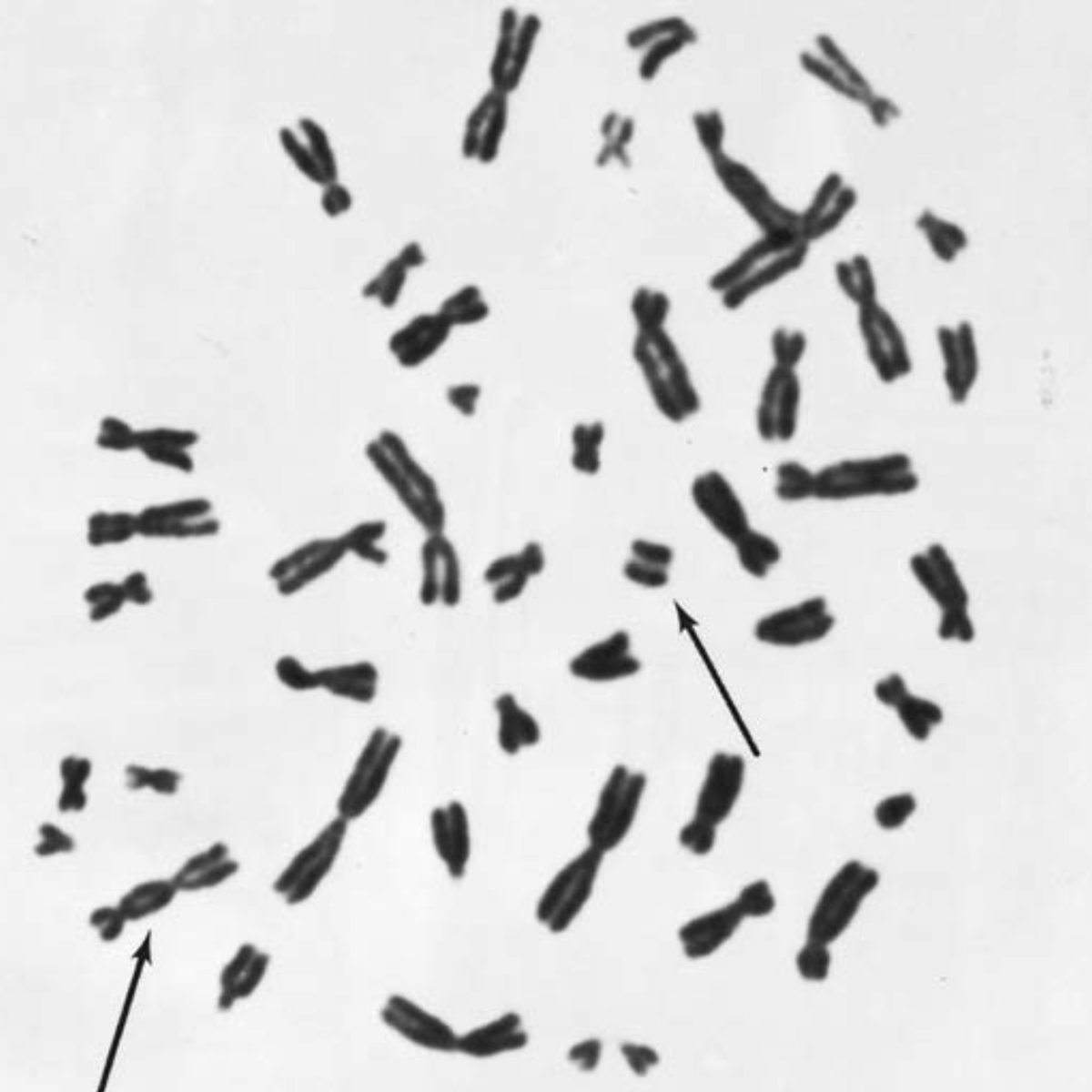
Damaged chromosomes after a radiation exposure.
– Radiobiology for the Radiobiologist, Eric Hall and Amato Giaccia
Chesal and Afshari want their phantoms to have the ability to be custom-tailored for any person, regardless of size, gender, body shape, or ethnicity.
“But how well we’ll be able to simulate and visualize stuff will still be based off of limitations in the original CT image or scan,” Chesal said.
Chesal knows a lot about imaging. In pursuit of her bachelor’s degree, she worked with Joyoni Dey, associate professor in the LSU Department of Physics & Astronomy. Dey holds two patents for innovations that have led to more detailed scans, while reducing dose, meaning amount of radiation, for the patient. The project that became Chesal’s honors thesis also relied on machine learning (a subfield of artificial intelligence) to improve the analysis of textures in scans to help predict tumor development.
Originally from Natchitoches, where her father manages a manufacturing plant for scroll compressors for air conditioners and her mother directs the office at the local Catholic basilica, Chesal first came to LSU in 2014 for a bachelor’s degree in physics with a minor in history, and then decided to stay for a master’s in medical physics—which now is looking more and more like it will pivot into a Ph.D.
“I have tried to leave LSU and Louisiana, but I keep being reeled back in,” Chesal said. “This is the best program in medical physics I’ve been able to find. So, here I am.”
A recent MBA graduate of LSU is Jonas Fontenot, originally from Crowley, Louisiana. As Chief Operations Officer and Chief of Physics at Mary Bird Perkins Cancer Center, Fontenot is at the forefront of the fight against cancer, especially due to advancements made through the renowned joint medical physics program and ongoing collaboration between LSU and the cancer care organization.
“Megan’s and Nousha’s work is key in the rapidly-growing science of precision medicine, which offers unprecedented opportunities to use increasingly detailed information to prevent, diagnose, and treat disease.”—Jonas Fontenot, Chief Operations Officer and Chief of Physics at Mary Bird Perkins Cancer Center
“Louisiana has some of the highest cancer mortality rates in the nation, so the need to advance care is critical in our state and beyond,” Fontenot said. “Megan’s and Nousha’s work is key in the rapidly-growing science of precision medicine, which offers unprecedented opportunities to use increasingly detailed information to prevent, diagnose, and treat disease. It’s the future of cancer care, and they are playing a critical role in advancing this area of study within radiation oncology, which will ultimately produce better outcomes and save more lives.”
The current phantoms used by the LSU SpaRTAN Lab are provided by researchers at Hanyang University Radiation Engineering Laboratory, HUREL.
Read more:
Going the Distance: Innovation in Radiation Research Takes LSU Medical Physics Student from Mary Bird Perkins Cancer Center to SpaceX (LSU Office of Research & Economic Development)
LSU Goes to the Moon (LSU Office of Research & Economic Development)